Primary surface measurement reported
Water contact angle (hydrogel wettability) was measured on GelMA and GelTA hydrogels prepared with and without EV incorporation.
Client Citation Analysis
Water contact angle (hydrogel wettability) was measured on GelMA and GelTA hydrogels prepared with and without EV incorporation.
The Dropometer is cited as a “Droplet Lab Tensiometer (Droplet Lab, Ontario, Canada)” used to inject 16 μl water droplets for contact angle measurement, with contact angles after 1 s analyzed by ImageJ software.
Contact angle was used to describe hydrogel hydrophilicity and to compare wettability between GelTA and GelTA‑EVs hydrogels. The text also references contact angle comparisons for GelMA and GelMA‑EVs in supplementary figures.
Contact angle in Fig. 5M was quantified by ImageJ (n = 3).
Water contact angle was measured after 1 s following deposition of a 16 μl water droplet on the hydrogel surface to characterize wettability.
Hydrogel microstructure and surface/topography context was reported using scanning electron microscopy (SEM) and atomic force microscopy (AFM) outputs presented alongside contact angle within the hydrogel characterization set.
Droplet Lab Tensiometer (Droplet Lab, Ontario, Canada)
ImageJ software
Hitachi S-4800 (Hitachi, Japan) scanning electron microscope (SEM)
AFM (ScanAsyst-Fluid + sharp-tipped cantilevers; DMT moduli evaluated computationally)
In the contact angle assay, hydrogels (GelMA or GelTA, prepared with or without EVs) were formed on glass using a 500 μm high and 10 mm diameter PDMS mold, swelled in DPBS for 3 h, and then dried with Kimwipes. A constant-volume water droplet (16 μl) was injected onto the gel surface using a Droplet Lab Tensiometer (Droplet Lab, Ontario, Canada), and the contact angle after 1 s was analyzed using ImageJ software.
These contact angle measurements were used to compare hydrogel wettability across formulations (including GelTA vs GelTA‑EVs) as part of the study’s hydrogel surface characterization.
GelTA and GelTA‑EVs hydrogels are described as having good hydrophilicity, with a contact angle around 50–70°, which the authors link to maximizing cell adhesion.
In Fig. 5M, GelTA and GelTA‑EVs hydrogels displayed similar contact angle, quantified by ImageJ (n = 3).
The text describes GelMA‑EVs as showing similar contact angle compared with pristine GelMA hydrogel and points to Figs. S6K and S6L.
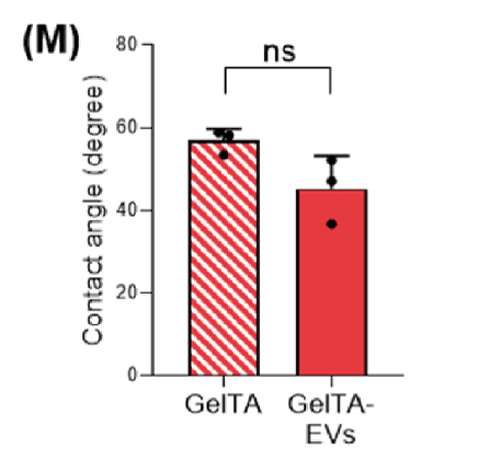
Shows GelTA and GelTA‑EVs hydrogels with similar contact angle, quantified by ImageJ (n = 3).
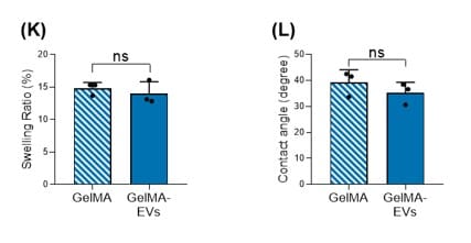
Referenced in the text for contact angle comparison between GelMA‑EVs and pristine GelMA hydrogels.
Within the hydrogel characterization workflow in this study, the contact angle assay provides a direct wettability readout used to describe surface hydrophilicity and to compare formulations with and without EV incorporation. The authors explicitly connect the reported contact angle range to cell adhesion considerations while presenting contact angle as part of the broader surface/structure characterization set for these biomaterial scaffolds.
The assay uses 16 μl water droplets and evaluates contact angle after 1 s, with angle analysis performed in ImageJ.
Hydrogels are swelled in DPBS for 3 h and then dried carefully with Kimwipes prior to droplet deposition.
Hydrogels are formed using a 500 μm high and 10 mm diameter PDMS mold and fixed on glass by placing a glass slide on top during preparation.
The study reports contact angle comparisons for GelTA vs GelTA‑EVs (Fig. 5M) and references GelMA vs GelMA‑EVs comparisons in supplementary figures.
The contact angle result in Fig. 5M is reported with n = 3.