Contents

Client Citation Analysis

Buffer Components Incorporate into the Framework of Polyserotonin Nanoparticles and Films during Synthesis

This study examines how synthesis-buffer chemistry affects polyserotonin (PSe) nanoparticles and films, using static contact angles on PSe films to calculate surface energy components.

At-a-Glance Summary

How the paper credits the instrument

The paper's complete attribution, Section 2.2: "Contact angles (Droplet Smart Tech, Markham, ON, Canada) and surface energy of PSe films synthesized in different buffers were also determined." No model number or software version is given. Angles were extracted by the authors' own code — "Contact angles were analyzed using a program that was written in-house" — and surface energy came from an OWRK calculation, so the instrument's contribution here is the drop imaging (0.3 Mp camera). [If the corporate-name decision above is approved, append:] Droplet Smart Tech is the corporate name under which these instruments were sold at the time of this 2022 study.

How the contact-angle data were used in the study

The static contact angles were used to compute the surface energy of PSe film-coated glass. The result was the same for every buffer tested: the paper reports that "for all buffer systems, the PSe film coating makes glass more hydrophilic," and gives no buffer-resolved contact-angle or surface-energy comparison in its main text.

What the Droplet Lab instrument did and did not do

It supplied static contact-angle images on polyserotonin films for three probe liquids, from which the authors' in-house code extracted angles and an OWRK calculation produced surface energies. It was not the primary measurement instrument; the paper's title finding — that buffer components incorporate into the polyserotonin framework — was established by FTIR and XPS and supported by DFT calculations, with nanomechanics and adhesion measured by a JPK Nanowizard 4 AFM. Contact-angle output appears in zero of the paper's four main-text figures and zero of its ten main-text panels; it is confined to Supplementary Figure S5 and Table S1.

Paper Details

Title
Buffer Components Incorporate into the Framework of Polyserotonin Nanoparticles and Films during Synthesis
Authors
Keuna Jeon; Justin Andrei Asuncion; Alexander Lucien Corbett; Tiange Yuan; Meera Patel; Nesha May Octavio Andoy; Christian Titus Kreis; Oleksandr Voznyy; Ruby May Arana Sullan
Journal
Nanomaterials
Year
2022
Volume
12
Pages / Article
2027
License
Creative Commons Attribution (CC BY) license (CC BY 4.0)
Funding & interests
Funded by the Canada Foundation for Innovation (Project 36544), the Ontario Ministry of Research, Innovation and Science (Project 36544), NSERC (RGPIN-2017-06522) and the Connaught Fund New Researcher Award; the first author held an NSERC PGS-D. All authors are at the University of Toronto Scarborough and the University of Toronto. The paper states: "The authors declare no conflict of interest. The funders had no role in the design of the study; in the collection, analyses, or interpretation of data; in the writing of the manuscript, or in the decision to publish the results." Droplet Smart Tech / Droplet Lab appears in no funding statement, no acknowledgement and no affiliation — the instrument was purchased and used as a lab tool.
Supplementary materials
Figures S1–S8 and Table S1, downloadable from the article page. The contact-angle images are Figure S5 ("Contact angle images of PSe films"); the film characterisation table, including thickness and surface-energy components, is Table S1. Both are supplementary — no contact-angle data appear in the four main-text figures.
9.2
Scopus metrics (Elsevier / Scopus rating 2024)
CiteScore 2024
Scopus metrics (Elsevier / Scopus rating 2024)
CiteScore subject ranks (CiteScore 2024)
  • Q1 - General Chemical Engineering (39/274)
  • Q1 - General Materials Science (82/460)
4.3
Journal Impact Factor (Clarivate JCR)
Journal Impact Factor (JCR 2024)
4.7
Journal Impact Factor (Clarivate JCR)
5-Year Impact Factor
Journal Impact Factor (Clarivate JCR)
JCR category rank
  • Q2 - Physics, Applied
  • Q2 - Chemistry, Multidisciplinary
  • Q2 - Materials Science, Multidisciplinary
  • Q2 - Nanoscience and Nanotechnology

What Was Measured

Primary surface / interfacial measurement

Static contact angles on PSe films using three probe liquids (milli-Q water, glycerol, diiodomethane), with surface energy (including dispersive and polar components) calculated using the OWRK model.

Supporting measurements

Chemical composition via FTIR spectroscopy and X-ray photoelectron spectroscopy (XPS) — the two techniques that established the paper's title finding; film thickness via AFM scratch tests; adhesion metrics from AFM force–distance curves; particle characterization via UV–Vis, TEM, DLS, zeta potential and EPR; binding energies via DFT calculation.

Instruments Mentioned

UV–Vis absorption spectra

Cary 60 UV–Vis (Agilent Technologies, Santa Clara, CA, USA)

TEM imaging

Hitachi H-7500 (with TiETEM version 5.2 software; Tokyo, Japan)

DLS (size distribution)

NanoBrook Omni (Brookhaven Instruments, Holtsville, NY, USA)

Zeta potential (PALS)

NanoBrook Omni (Brookhaven Instruments, Holtsville, NY, USA)

EPR (free radical centers)

Bruker X-band CW EMX EPR spectrometer (10” electromagnet; ER4123D resonator)

Contact angles

Droplet Smart Tech (Markham, ON, Canada); camera 0.3 Mp; angles extracted with an in-house written program; surface energy calculated with the OWRK model

AFM (quantitative imaging; nanomechanics)

Nanowizard 4 (JPK Instruments, Berlin, Germany)

Film thickness processing

JPK Image Processing software

DFT calculations

CP2K

FTIR

Fourier-transform infrared spectroscopy — produced Figure 2A, the S=O stretching peaks at 1369 and 1056 cm⁻¹ that identify HEPES inside the nanoparticle framework. The paper names no FTIR instrument, manufacturer or model in Section 2.2.

XPS

X-ray photoelectron spectroscopy — produced Figure 2B and Figure S2, including the S2p sulfur peak that confirms HEPES incorporation. No instrument is named in the Methods; XPS is credited only in the Acknowledgements, to "Rana N.S. Sodhi from the Ontario Centre for Characterization of Advanced Materials (OCCAM)".

Role of the Droplet Lab instrument

Static contact angles were measured using an instrument the paper attributes only as "Droplet Smart Tech, Markham, ON, Canada". Section 2.2: "20 µL of pure diiodomethane, glycerol, and milli-Q water were manually dropped onto the films and imaged with a camera (0.3 Mp) to estimate the angles of each drop. Contact angles were analyzed using a program that was written in-house, and the surface energy was calculated using the Owens–Wendt–Rabel–Kaelbel (OWRK) model." The instrument supplied the drop imaging; the angle values were produced by the authors' own code, and the surface energies are OWRK model outputs, not instrument readings.

These contact-angle-derived surface-energy results establish a single common result across all four synthesis buffers: "for all buffer systems, the PSe film coating makes glass more hydrophilic, decreasing the water contact angle by over 15° and increasing the surface energy of glass from ~47 to ~61 mJ/m²." The paper reports no buffer-to-buffer difference in contact angle or surface energy in its main text, and its Conclusions do not cite either. What did discriminate between the buffers were AFM adhesion force, work of adhesion, film thickness and zeta potential not wettability.

Method Snapshot

Notes: All four rows carry identical measurement conditions and identical data locations because the paper reports a single common outcome for every buffer — "for all buffer systems, the PSe film coating makes glass more hydrophilic, decreasing the water contact angle by over 15° and increasing the surface energy of glass from ~47 to ~61 mJ/m²". Individual per-buffer contact angles are not given in the main text; they are in Table S1. The paper states no replicate count, temperature or relative humidity for these measurements, drops were placed manually at 20 µL, and only static angles were recorded.

Film system (as prepared in the study) Buffer used during synthesis Surface outputs derived from droplets Instruments Conditions (as reported) Data location (Supplementary)
PSe film-coated glass coverslips Tris Static contact angles (water, glycerol, diiodomethane); surface energy (dispersive + polar) via OWRK Droplet Smart Tech; camera (0.3 Mp); in-house program 20 µL droplets; liquids manually dropped onto films Figure S5; Table S1
PSe film-coated glass coverslips DEA Static contact angles (water, glycerol, diiodomethane); surface energy (dispersive + polar) via OWRK Droplet Smart Tech; camera (0.3 Mp); in-house program 20 µL droplets; liquids manually dropped onto films Figure S5; Table S1
PSe film-coated glass coverslips Bicine Static contact angles (water, glycerol, diiodomethane); surface energy (dispersive + polar) via OWRK Droplet Smart Tech; camera (0.3 Mp); in-house program 20 µL droplets; liquids manually dropped onto films Figure S5; Table S1
PSe film-coated glass coverslips HEPES Static contact angles (water, glycerol, diiodomethane); surface energy (dispersive + polar) via OWRK Droplet Smart Tech; camera (0.3 Mp); in-house program 20 µL droplets; liquids manually dropped onto films Figure S5; Table S1

Key Findings

PSe film coating increases hydrophilicity

The paper reports, in Section 3.4: "for all buffer systems, the PSe film coating makes glass more hydrophilic, decreasing the water contact angle by over 15°." The effect is reported as common to all four synthesis buffers; no per-buffer water contact angle is given in the main text.

Surface energy increases upon PSe film coating

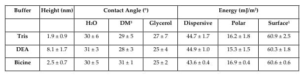
Surface energy of glass increases from ~47 to ~61 mJ/m² when coated with PSe films (Table S1). These are OWRK model outputs computed from the three static contact angles, not direct instrument readings; the paper does not publish the dispersive/polar reference values used for water, glycerol and diiodomethane, so the calculation cannot be reproduced from the article alone.

Dispersive contribution dominates the surface-energy increase

The paper notes that "although the PSe film increased the overall surface energy of the glass, there is a higher contribution from the increase in the dispersive energy component than the polar energy component (Table S1)." Its stated significance is a contrast with polydopamine: "This is significantly different from polydopamine films, where an increase in surface energy was largely driven by increasing polar energy component, independent of the substrate used." The polydopamine comparison is drawn from prior literature (Bourmaud et al. 2009; Jiang et al. 2011), not from measurements made in this study.

HEPES incorporates into the polyserotonin framework (FTIR + XPS)

The paper's title finding. Nanoparticles synthesised in HEPES showed FTIR peaks at 1369, 1056 and 838 cm⁻¹ absent from the other three buffers; the first two are assigned to S=O stretching from the sulfur group of HEPES. High-resolution XPS confirmed a S2p sulfur peak in the HEPES particles only. The paper is precise about the limit of this evidence: "HEPES, out of the four buffer components, has a unique sulfur atom that allows this difference to be identified." Direct spectroscopic evidence therefore exists for one buffer of four; for the other three the authors infer — "we posit that other buffer components are also likely to become trapped". The XPS survey spectrum showed no difference in oxygen, nitrogen or carbon composition between buffers.

DFT binding energies support incorporation, at monomer level only

All four buffer components bind favourably to a serotonin monomer at the pH 9 synthesis condition: Bicine −3.745 eV, HEPES −3.281 eV, Tris −1.640 eV, DEA −1.469 eV. Binding is weaker at pH 7 for all four, and the HEPES/Bicine order reverses (HEPES −2.638 eV, Bicine −1.733 eV). The calculation's limits are stated by the authors: "we used the structure of serotonin monomer since the mechanism of polyserotonin formation is currently unknown, and the structures of the early intermediates of nanoparticle formation have not been investigated." They close by noting the work is unfinished: "We are currently investigating whether the buffer components interact with early intermediates of polyserotonin formation, which could then lead to physical entrapment during nanoparticle growth."

What buffer choice did not change

Three null results, all stated by the authors. Elasticity: "we did not observe a significant difference in the Young's modulus of the particles as a function of the buffer system" — apparent Young's modulus was ~400 MPa across all four. Radical species: "an EPR analysis showed no difference in g-values (2.0038–2.0042) across all four buffer types further indicating the formation of the same semiquinone radical species, regardless of the buffer used." Bulk elemental composition: the XPS survey spectrum "showed no difference in the atomic composition of oxygen, nitrogen, and carbon". Wettability belongs on this list — the contact-angle result was reported as common to all four buffers.

Adhesion and film thickness — not wettability — separated the buffers (JPK Nanowizard 4 AFM)

Against an APTES-functionalised (positively charged) AFM tip, adhesion force on PSe films was 1.6 ± 0.3 nN for Tris and 1.8 ± 0.5 nN for HEPES, versus 1.2 ± 0.2 nN for both DEA and Bicine, and 0.4 ± 0.1 nN for bare glass (n = 153). Work of adhesion followed the same order: Tris and HEPES 0.07 fJ, DEA 0.04, Bicine 0.03, glass 0.01 (n = 145, one-way ANOVA, p ≤ 0.001). Film thickness ranged from 1.9 ± 0.9 nm in Tris to 9.1 ± 2.1 nm in HEPES. These, plus zeta potential, are the film properties the paper's Conclusions cite as evidence that buffer choice matters — contact angle and surface energy are not mentioned in the Conclusions.

Figures & Visuals

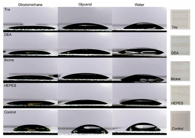
Supplementary Figure S5 — contact angle images of PSe films

Supplementary Figure S5 — contact angle images of PSe films

Shows the static contact angle images on PSe films for the three probe liquids used in the OWRK surface-energy analysis. The paper's own caption for this panel is simply "Contact angle images of PSe films".

Supplementary Table S1 — characterisation of PSe films on glass

Supplementary Table S1 — characterisation of PSe films on glass

Summarises film properties in the discussion of contact-angle changes and the resulting surface energy (dispersive and polar components). The same table also carries the AFM film-thickness values that do vary by buffer — 1.9 ± 0.9 nm for Tris up to 9.1 ± 2.1 nm for HEPES.

Why It Matters

Within the study's buffer-dependent synthesis framework, the contact angle measurements answered a specific question: does the choice of synthesis buffer change how a polyserotonin film wets? The answer was no. The paper reports one outcome for every buffer — "for all buffer systems, the PSe film coating makes glass more hydrophilic, decreasing the water contact angle by over 15°" — and the three-liquid protocol allowed that to be expressed as surface-energy components rather than a single angle. Knowing which property is insensitive to a synthesis variable is what lets the next lab stop controlling for it.

The surface-energy decomposition (dispersive vs polar) served one specific argument in the paper: that polyserotonin films behave differently from polydopamine films, where "an increase in surface energy was largely driven by increasing polar energy component" in the prior literature. That is the only use the paper makes of the decomposition. The buffer-dependent conclusions — that Tris and HEPES yield the most negative surfaces, that Tris gives the thinnest films and most monodisperse particles — rest on zeta potential, AFM adhesion and AFM thickness, not on wettability.

This is an independent citation. The work was funded by the Canada Foundation for Innovation, the Ontario Ministry of Research, Innovation and Science, NSERC and the Connaught Fund, carried out at the University of Toronto Scarborough, and the authors declare no conflict of interest and record that "the funders had no role in the design of the study; in the collection, analyses, or interpretation of data; in the writing of the manuscript, or in the decision to publish the results." Droplet Smart Tech appears nowhere in the funding, acknowledgements or affiliations — it appears once, in a Methods parenthesis, as a tool the lab bought and used.

Practical Takeaways

Three-liquid contact-angle workflow

Static contact angles were collected using 20 µL drops of milli-Q water, glycerol, and diiodomethane, with camera-based imaging and in-house angle extraction feeding an OWRK surface-energy calculation.

Hydrophilicity shift after coating

The reported outcome for PSe film-coated glass is a reduction in water contact angle by over 15° — and the paper reports this "for all buffer systems", with no per-buffer difference given in the main text.

Surface energy rises with coating

The reported total surface energy increases from ~47 to ~61 mJ/m² when glass is coated with PSe films (Table S1) — an OWRK calculation from the three measured angles, not a directly measured quantity.

Dispersive component drives the increase

The study attributes more of the surface-energy increase to the dispersive component than to the polar component (Table S1) — the reverse of what the polydopamine literature reports, which is why the authors flag it.

Read these angles as a screening measurement

The protocol was 20 µL drops placed manually and imaged with a 0.3 Mp camera, with angles extracted by unpublished in-house code. The paper reports no replicate count, no temperature and no humidity for these measurements, and no advancing/receding angles or hysteresis. A 20 µL water drop is well above the capillary length and is gravity-flattened, so absolute values will sit below what a smaller drop would give. That is adequate for the comparison the paper actually makes — coated versus uncoated glass, a >15° shift — and not a basis for treating the individual surface energies as precise. If you are reproducing this work, dispense smaller drops, state n, and record the temperature.