Contents

Client Citation Analysis

A Mechanistic, Architecture-Dependent Study Combining Experiments and Molecular Dynamics to Explain AMP Release from GO–PEI Coatings

This study compares layered PEI+GO and embedded PEI/GO antimicrobial coatings on urinary catheters, with static sessile-drop contact-angle measurements used to track coating-induced wettability alongside morphology, molecular dynamics, and antibacterial performance.

At-a-Glance Summary

How the paper credits the instrument

Section 2.5, Characterization Techniques: "Wettability of coated UC samples was assessed via static sessile drop (10 µL, n = 5) using a Droplet Lab system (Brampton, ON, Canada)." That is the paper's only mention of the instrument. No product or model name is given, no software version, and no drop-fitting method (polynomial or Young–Laplace) is stated.

How the contact-angle data were used in the study

The contact-angle data were used to compare the control, PEI+GO and PEI/GO surfaces and to show that the coating processes increased hydrophilicity together with surface roughness. The paper's own summary of this result is one sentence: "both samples exhibited increased surface roughness and hydrophilicity as a result of the coating processes (Table 1)." The data are not used again anywhere in the Results, Discussion or Conclusions. These surface-property results sat alongside FTIR, Raman, microscopy, molecular dynamics, and antibacterial assays in the architecture-dependent comparison.

Replication / reliability statement

Wettability was assessed by static sessile drop using 10 µL droplets with n = 5. The paper states no measurement temperature, no humidity, no equilibration or image-capture timing, and does not say whether the ± 1.0° in Table 1 is a standard deviation or a standard error. No advancing/receding angles or hysteresis were measured, and no drop-fitting model is named.

What the Droplet Lab instrument did and did not do

It measured static water contact angle on three bare surfaces — uncoated control, PEI+GO and PEI/GO — producing Figure 3B and one column of Table 1, 1 of the paper's 5 figures. It was not the primary measurement instrument; the paper's principal outcomes were produced by NAMD 2.13 molecular dynamics simulations and agar diffusion assays. No contact angle was measured on any peptide-loaded coating, and the instrument did not measure peptide release.

Paper Details

Title
A Mechanistic, Architecture-Dependent Study Combining Experiments and Molecular Dynamics to Explain AMP Release from GO–PEI Coatings.
Authors
Adriana de América; María José Fritte; Paola Alarcón; Karel Mena-Ulecia; Gonzalo Recio-Sánchez; Klaus Rischka; Marcos Rocha Diniz Silva; Matheus Santos Dias; Camila Marchetti Maroneze; Cecilia de Carvalho Castro Silva; Jacobo Hernandez-Montelongo.
Journal
Bioengineering
Year
2026
Volume
13
Issue
3
Pages / Article
341
License
CC BY
Funding & interests
Funded by FONDECYT–Chile (No. 1230553), CAPES, MackPesquisa, CNPq (Nos. 408248/2023-8, 313091/2022-6), INCT NanoVida (No. 406079/2022-6) and Finep (Nos. 1151/22, 1755/22); supercomputing infrastructure provided by NLHPC (CCSS210001). Authors are at Universidad Católica de Temuco and Universidad San Sebastián (Chile), Fraunhofer IFAM (Germany), and Mackenzie Presbyterian University / MackGraphe (Brazil). "The authors declare no conflicts of interest." Droplet Lab appears in no funding, acknowledgement, affiliation or conflict-of-interest statement — the instrument was purchased and used as a lab tool.
Data availability
"The dataset is available on reasonable request from the authors." No public repository deposit; the raw contact-angle images and per-replicate values are not downloadable.
5.3
Scopus metrics (Elsevier / Scopus rating 2024)
CiteScore 2024
Q3 - Bioengineering
Scopus metrics (Elsevier / Scopus rating 2024)
CiteScore subject ranks (CiteScore 2024)
0.735
Scopus metrics (Elsevier / Scopus rating 2024)
SJR 2024
3.7
Journal Impact Factor (Clarivate JCR)
Journal Impact Factor (JCR 2024)
3.9
Journal Impact Factor (Clarivate JCR)
5-Year Impact Factor
Not stated. (JCR category ranking: Q2, Engineering — Biomedical.)
Journal Impact Factor (Clarivate JCR)
JCR category rank

What Was Measured

Primary surface / interfacial measurement

Static water contact angles were determined for control, PEI+GO and PEI/GO catheter samples, yielding values of 98.3 ± 1.0°, 76.4 ± 1.0° and 75.5 ± 1.0° respectively — all three measured on bare coatings before antimicrobial peptide loading. The two coated architectures differ by 0.9°, inside the reported uncertainty; the coating step is what the measurement resolves, not the choice between architectures.

Supporting measurements

Zeta potential, surface roughness, FTIR, Raman spectroscopy, optical microscopy, VP-SEM and FEG-SEM characterised the coatings. The paper's two principal outputs are not supporting measurements: 50 ns NAMD 2.13 molecular dynamics simulations of the GO–AMP and PEI–AMP complexes (Figure 5A,B) and agar diffusion antibacterial assays against E. coli and E. faecalis (Figure 5C) together produce the mechanistic conclusion the paper's title announces.

Instruments Mentioned

Wettability / water contact angle

Droplet Lab system (Brampton, ON, Canada)

Zeta potential

Zetasizer Advance (Malvern; Worcestershire, UK)

Surface roughness

PCE-RT 2300 tester (PCE Instruments; Southampton, UK)
FTIR: 4600, Jasco (Tokyo, Japan)

Raman spectroscopy

Dual Hound, Unchained Labs (Pleasanton, CA, USA)

Optical microscopy

Eclipse E200, Nikon (Tokyo, Japan)

VP-SEM

SU-3500, Hitachi (Tokyo, Japan)

FEG-SEM

JSM-7800, Jeol (Tokyo, Japan)

MALDI-ToF MS

Autoflex Speed, Bruker (Billerica, MA, USA)

Molecular dynamics engine

NAMD 2.13 — 50 ns production runs at 300 K, 1 fs step, 12 Å cutoff, PME, TIP3P solvation; CHARMM36 / CGenFF / SwissParam force fields

DFT geometry optimisation

Orca (B3LYP/DEF2-TZVPP), GO sheet built in VMD and Chemcraft

Plasma surface activation

Harrick Plasma (Ithaca, NY, USA) — 15 min, < 0.2 mmHg, 18 W

Peptide synthesis

Liberty Blue microwave-assisted synthesizer, CEM (Kamp-Lintfort, Germany)

Statistics

OriginLab v.2022b — ANOVA with Tukey post hoc

Role of the Droplet Lab instrument

The Droplet Lab instrument appears once in the characterization workflow, in Section 2.5: "Wettability of coated UC samples was assessed via static sessile drop (10 µL, n = 5) using a Droplet Lab system (Brampton, ON, Canada)." The paper names no model and no software version. The resulting water contact angles are reported for the control, PEI+GO and PEI/GO samples — the three bare surfaces, measured before either antimicrobial peptide was loaded.

In the study's comparison of coating architectures, the wettability results showed that both GO-based coatings shifted the catheter surface toward higher hydrophilicity — from 98.3 ± 1.0° to 76.4 ± 1.0° and 75.5 ± 1.0°. What they did not do was separate the two architectures from each other: those two values differ by 0.9°, inside the reported uncertainty, for coatings that differ by 570 nm in thickness and by the paper's widest antibacterial margin. The layered and embedded structures were separated by morphology, thickness, simulated interaction energetics and antibacterial response, and the paper's explanation of peptide release invokes none of them through wettability.

Key Findings

Coating lowered water contact angle

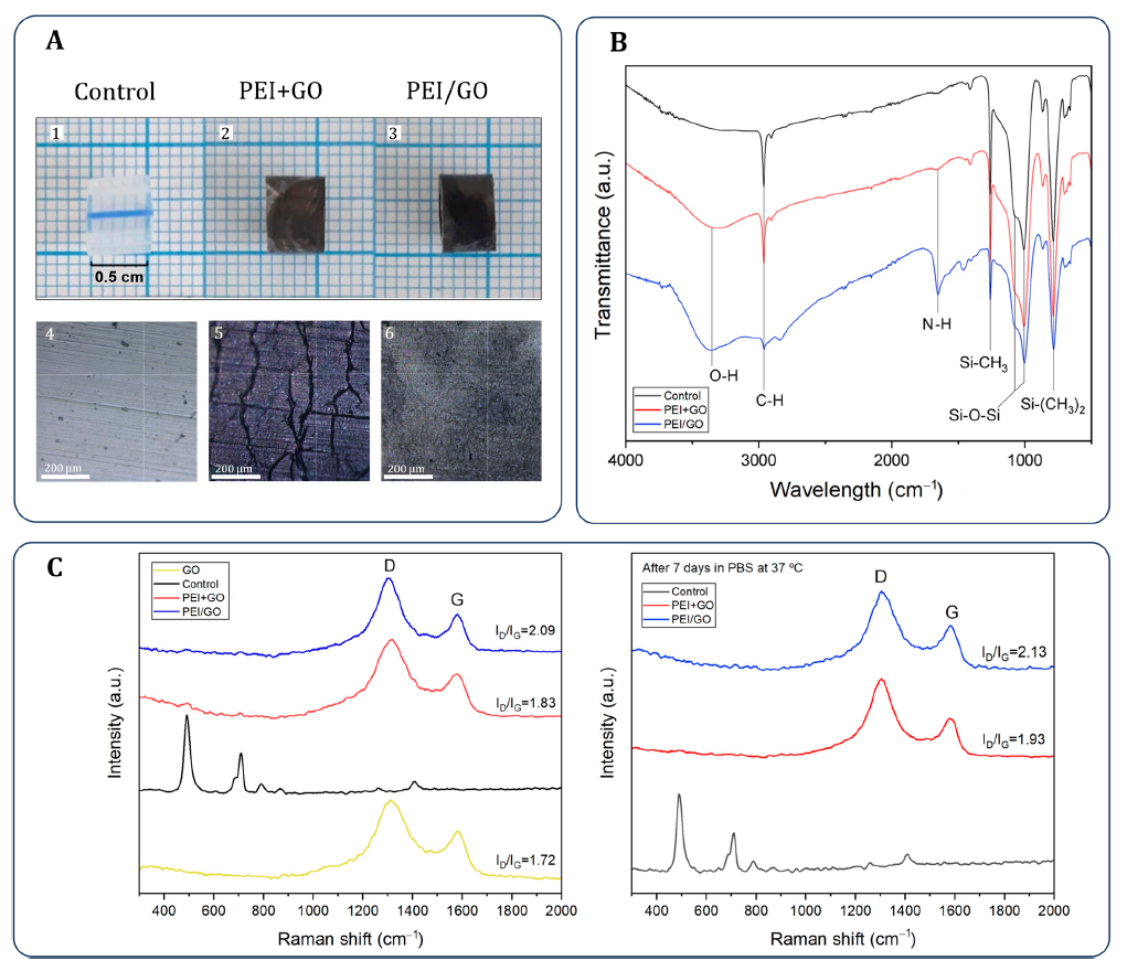
The control catheter showed a water contact angle of 98.3 ± 1.0°, while PEI+GO and PEI/GO measured 76.4 ± 1.0° and 75.5 ± 1.0° respectively. The authors' own summary: "both samples exhibited increased surface roughness and hydrophilicity as a result of the coating processes."

Architecture changed surface morphology and thickness

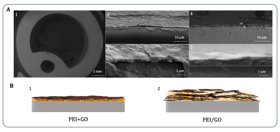
Microscopy described PEI+GO as porous and cracked, whereas PEI/GO appeared smoother. Cross-sectional SEM gave an average thickness of 320 ± 33 nm for PEI+GO and 890 ± 115 nm for PEI/GO — the paper notes the embedded coating is "nearly three times thicker".

PEI–peptide complexes were more stable in simulation

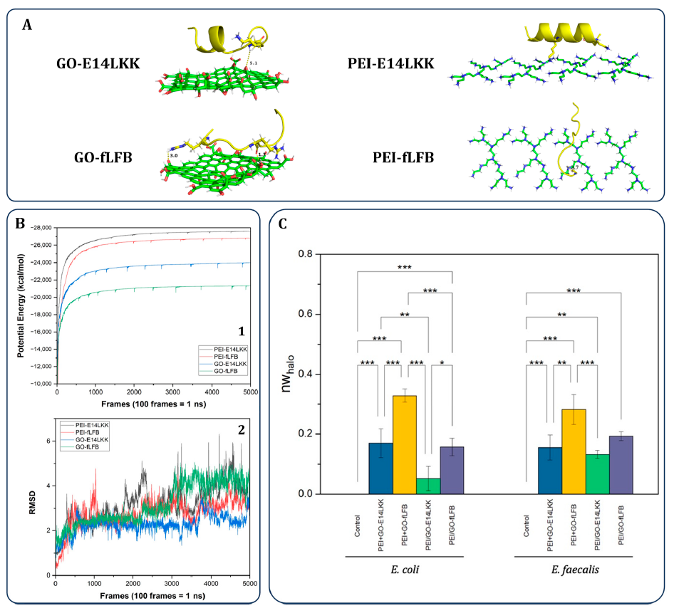
Potential-energy profiles stabilized from about 8 ns onward, and the most negative energies corresponded to the PEI complexes with E14LKK and fLFB. The paper's inference: "these complexes remain more stable over time, and therefore, the peptides will be released less into the PEI matrix than GO." These are NAMD 2.13 simulation outputs over 50 ns, not measurements.

GO–fLFB showed the highest mobility

The GO–fLFB complex maintained the highest potential energy and showed the greatest RMSD fluctuations among the four modeled complexes — all four had mean RMSD above 3 Å. The authors conclude: "fLFB will be released more readily in this matrix." Both quantities are simulation outputs; the paper notes the simulations modelled the GO–AMP and PEI–AMP complexes individually, "rather than the full coating architectures".

Layered PEI+GO–fLFB produced the strongest antibacterial response

Against both E. coli and E. faecalis, the PEI+GO–fLFB coating produced the largest inhibition zones (agar diffusion, Mueller–Hinton, 0.5 McFarland, 35 °C for 24 h, n = 3). The paper attributes this to "(i) the weaker interaction between GO and fLFB, which facilitates peptide release... and (ii) the coating architecture", and records that PEI/GO–E14LKK was the weakest of all samples tested. The authors caveat the method itself: "antimicrobial peptides (AMPs) generally exhibit limited diffusivity in agar-based systems."

Architecture and interaction strength were interpreted together

The study's conclusion is that coating configuration and GO–AMP interaction strength together dictated antimicrobial performance on urinary catheters; in the tested set, PEI+GO outperformed PEI/GO. The paper's own wording: "These findings underscore the importance of both coating architecture and AMPs–GO interaction strength in dictating antimicrobial performance."

Contact angle did not distinguish the two architectures

PEI+GO measured 76.4 ± 1.0° and PEI/GO 75.5 ± 1.0° — a 0.9° gap inside the stated uncertainty, with no statistical test reported between them. Over the same two coatings, cross-sectional SEM found 320 ± 33 nm versus 890 ± 115 nm, Raman found I(D)/I(G) of 1.83 versus 2.09, and the agar assays separated PEI+GO–fLFB (largest inhibition zones) from PEI/GO–E14LKK (weakest of every sample tested). Wettability resolves the coating step; it does not resolve the architecture, and the paper never claims it does — its explanation of the difference is "the weaker interaction between GO and fLFB, which facilitates peptide release" and "screening of GO's functional groups" in the embedded structure.

No release curve was measured — release was inferred

The paper contains no UV-vis, HPLC or any other time-resolved measurement of peptide release. Its abstract describes the agar assays as "a comparative indicator of peptide release from surface-bound coatings", and the release mechanism is reconstructed from simulated potential energy and RMSD together with inhibition-zone size. The authors are explicit that this is mechanistic groundwork: "Rather than targeting immediate clinical translation, this work provides mechanistic insight into how GO–polymer architecture modulates antimicrobial peptide availability."

Figures & Visuals

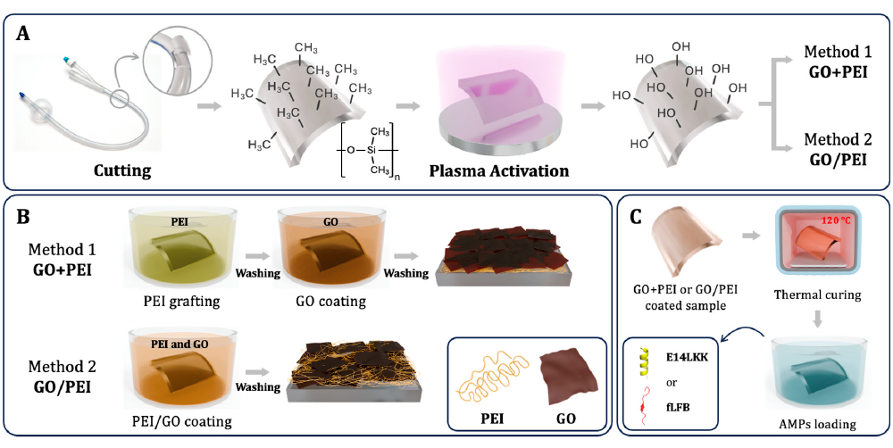
Figure 1 — coating process schematic

Figure 1 — coating process schematic

Figure 1. This schematic lays out the plasma pretreatment (15 min, 18 W), the PEI+GO and PEI/GO coating routes, and the thermal curing (120 °C, 1 h) and AMP loading steps used in the overall study design. The authors flag it as non-literal: "The scheme is illustrative; chemical changes are idealized," and "All assays used cylindrical catheter sections (0.5 cm); the rectangular shape in Figure 1 is a schematic reference only."

What it shows

What it shows

Figure 3B and Table 1 present the wettability and roughness comparison for the control, PEI+GO and PEI/GO samples. The contact angles come from the Droplet Lab sessile-drop system; the R(RMS) roughness values in the same panel (0.16, 0.25 and 0.28 ± 0.03 µm) come from a PCE-RT 2300 tester measured per ISO 4287. This is the only panel in the paper containing Droplet Lab data.

What it shows

What it shows

Cross-sectional SEM in Figure 4 shows the layered PEI+GO structure (320 ± 33 nm, folded and corrugated) and the embedded PEI/GO structure (890 ± 115 nm, smoother and more homogeneous). This is where the two architectures visibly diverge — a 570 nm thickness difference that the contact-angle measurement in Figure 3B does not register, since the two coatings' angles differ by 0.9°.

What it shows

What it shows

Figure 5 combines molecular dynamics outputs (5A: the four complexes; 5B(1): 50 ns potential-energy profiles; 5B(2): RMSD) with antibacterial data (5C: inhibition zones vs E. coli and E. faecalis, mean ± SD, n = 3, ANOVA with Tukey) to interpret why the PEI+GO–fLFB system showed the strongest activity. The simulations modelled GO–AMP and PEI–AMP complexes individually rather than the assembled coatings; the authors list extending them "to encompass the complete coating architectures" as future work.

Why It Matters

For this urinary-catheter coating study, the Droplet Lab contact-angle data establish the surface-state shift that follows GO-based coating deposition: the uncoated silicone control sat at 98.3 ± 1.0°, both coated surfaces at roughly 76°. Wettability is the paper's direct readout for whether a coating is present and has changed the interface. It is not a readout for which architecture was used — PEI+GO and PEI/GO returned 76.4 ± 1.0° and 75.5 ± 1.0°, and the paper draws no distinction between them on this basis.

Those wettability results matter because they confirm, cheaply and quickly, that the coating deposited and altered the surface — the check that has to pass before any of the downstream work means anything. The explanation of peptide availability itself rests elsewhere: the authors attribute the PEI+GO–fLFB result to "the weaker interaction between GO and fLFB, which facilitates peptide release", supported by simulated potential-energy and RMSD profiles, and to the embedded architecture "screening of GO's functional groups". Contact angle is not part of that causal chain, and the paper never places it there.

What this study does not establish, in the authors' own terms: it is not a clinical result — "this study does not aim to deliver a clinically validated catheter coating". Peptide release was never measured directly; agar inhibition zones stand in for it, and the authors note that "antimicrobial peptides (AMPs) generally exhibit limited diffusivity in agar-based systems". The simulations modelled isolated GO–AMP and PEI–AMP complexes, "rather than the full coating architectures". Coating durability was probed only by Raman after 7 days in PBS at 37 °C, with no mechanical or flow challenge. The authors' own next steps are "complementary antibiofilm assays" and expanded simulations covering the complete architectures.

Practical Takeaways

Use wettability as an architecture check

In this paper, sessile-drop contact angle clearly separated the uncoated catheter from both GO-coated surfaces, making it a straightforward readout of coating-induced surface change.

Expect morphology to matter beyond angle alone

PEI+GO and PEI/GO had very similar contact angles, yet they differed strongly in morphology and thickness, which became important in the later release and antibacterial interpretation.

Pair contact angle with structural measurements

The authors did not rely on wettability by itself; they interpreted it alongside roughness, FTIR, Raman, and SEM to characterize the coated surfaces.

Connect surface data to functional outputs

The study ran three layers of evidence — surface characterization, peptide-interaction simulations and antibacterial assays — but they do different jobs. Characterization establishes that the two architectures were built as intended; the simulations and assays explain the performance difference between them. Use surface measurements to verify the sample is what you think it is, then expect the mechanism to come from elsewhere.

Measure the surface in the state you are testing

Every contact angle in this paper was measured on a bare coating. All four functional systems — PEI+GO–E14LKK, PEI+GO–fLFB, PEI/GO–E14LKK, PEI/GO–fLFB — carry an adsorbed peptide layer applied at 0.5 mg/mL for 1 h, and none was measured. Peptide adsorption is exactly the kind of change that shifts wettability, so if you are correlating surface state with release behaviour, measure after loading as well as before. It is a cheap addition to a protocol and it closes the gap between what was characterised and what was tested.